
Aortic Valve Diseases and Surgery
What does the aortic valve do? How does it work?
A healthy heart beats approximately 70-80 times per minute and about 100,000 times per day. Thus, it pumps almost 300 liters of blood per hour throughout the body. A normal heart has four cavities. The two upper cavities are called the right and left atria, and the two lower cavities are called the right and left ventricles. The job of the heart is to send oxygen-rich blood throughout the body. Blood passes through the cavities of the heart and the four heart valves until released into the body. The blood, purified and oxygenated in the lungs, passes through these four cavities and is finally released from the aortic valve into the main artery, the “Aorta,” and from there is sent throughout the body. The aortic valve is between the left ventricle and the main artery (main artery). It ensures the pumping of blood from the heart to all organs.
What are aortic valve diseases?
Problems with the aortic valve are usually caused by underlying diseases, such as aortic stenosis or aortic valve regurgitation. Aortic valve stenosis makes it difficult for blood to pass through the body, and insufficiency causes blood to flow back from the aorta, that is, to the heart.
In a healthy heart, the aortic valve consists of three leaflets. The fact that the aortic valve is appears with two leaflets is called a bicuspid aortic valve and is the most common condition among congenital heart defects. Although it usually does not cause any signs or symptoms in its early stages, it can cause complications such as aortic valve disease (aortic stenosis, aortic regurgitation), aortic aneurysm (enlarged aorta), aortic dissection (ruptured aorta), and infective endocarditis.
What are the symptoms of aortic valve disease?
Complaints such as chest pain, tightness, feeling faint and faint after tiring work, imbalance, weakness, shortness of breath, and palpitations may be symptoms of aortic valve disease and should raise red flags.
How to diagnose aortic valve diseases?
The examination is carried out in connection with the patient’s complaints.
The final diagnosis is made when the doctor hears a heart murmur and after preliminary tests, echocardiography, and, if necessary, cardiac catheterization and angiography.
What are the treatments for aortic valve disease?
Treatment of all valve diseases consists of replacement (change) or reconstruction (repair).
What is aortic stenosis?
The aortic valve becomes calcified and distorted, causing it to become severely narrowed and unable to open normally. This means that the heart has to pass through a very narrow valve as it pumps blood throughout the body. Therefore, the heart muscle experiences great tension and stress. This can cause the heart muscle to thicken over time and, at a later stage, become weaker and insufficient. Heart failure is a condition that impairs a person’s health and causes limited movement. Persistence of valvular stenosis and heart failure represents a life-threatening risk situation.
What causes aortic stenosis?
It often develops due to age-related wear and tear on the aortic valve and the deposition of calcified deposits that narrow the valve and limit its movement. The development of the disease is facilitated by rheumatism, radiation therapy, and high cholesterol.
How to treat aortic stenosis?
Medication makes it impossible to correct the calcified stenosis that has formed in the aortic valve over the years. However, supportive drug treatment is provided for heart failure, arrhythmia and the risk of blood clots due to aortic valve disease. Treatment of aortic stenosis: It is performed surgically and through a catheter.
What are the treatments for aortic valve disease?
Today, aortic valve replacement is performed using 3 different methods:
●Open heart surgery, large incision (sternotomy)
●Closed aortic valve replacement or repair using a small incision in the armpit or chest (using a 3 or 4 cm long skin incision)
●The TAVI method (transfemoral aortic valve implantation)
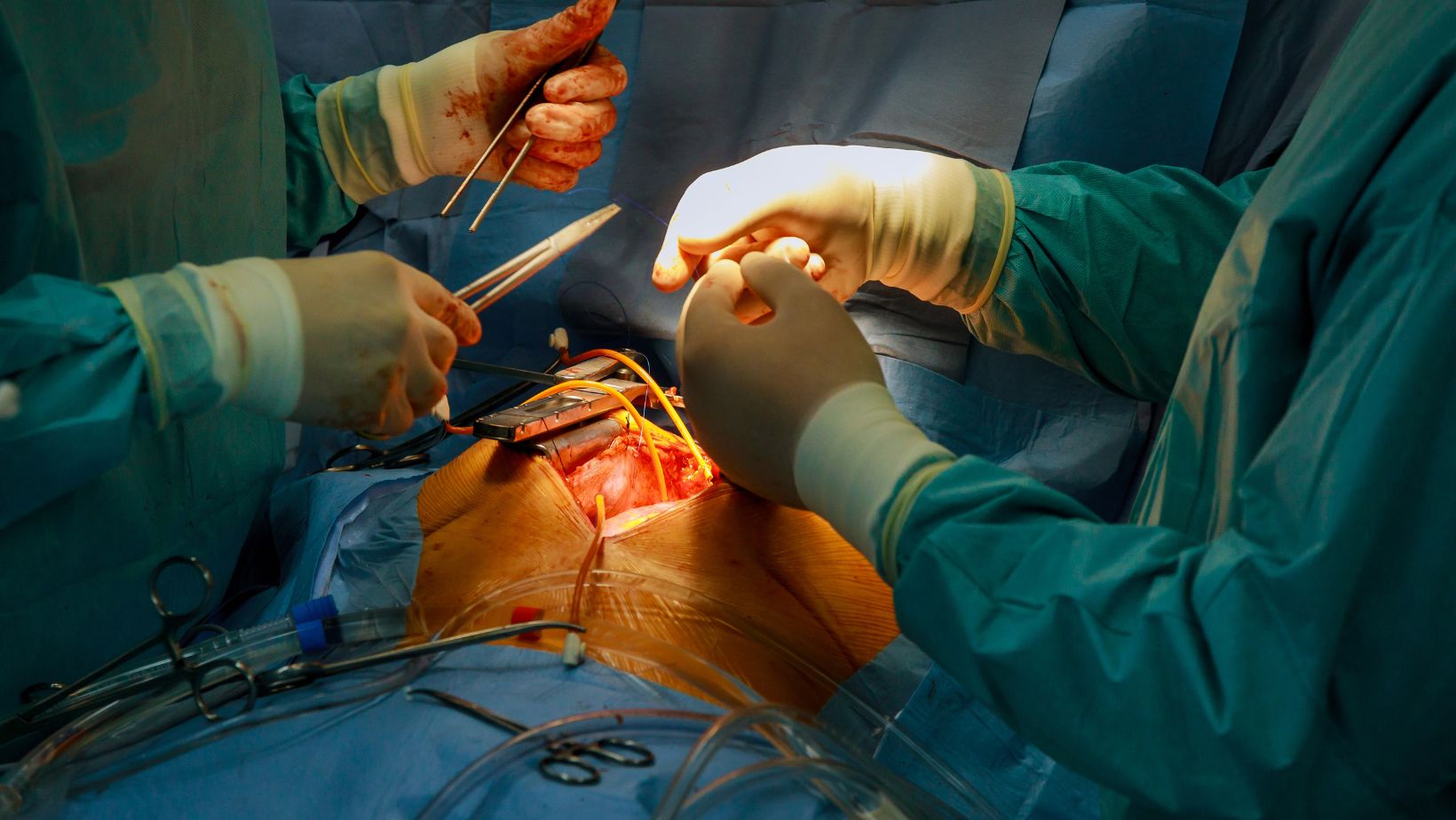
Method 1: Classic open heart surgery method.
Method 2 Closed heart surgery with a small incision
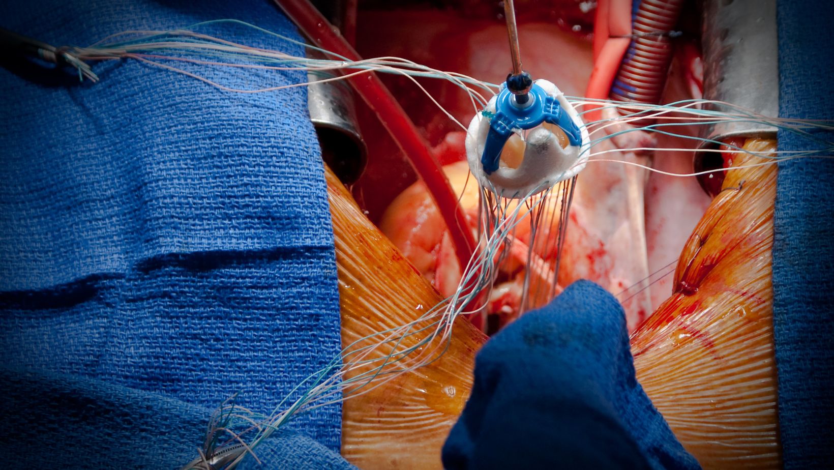
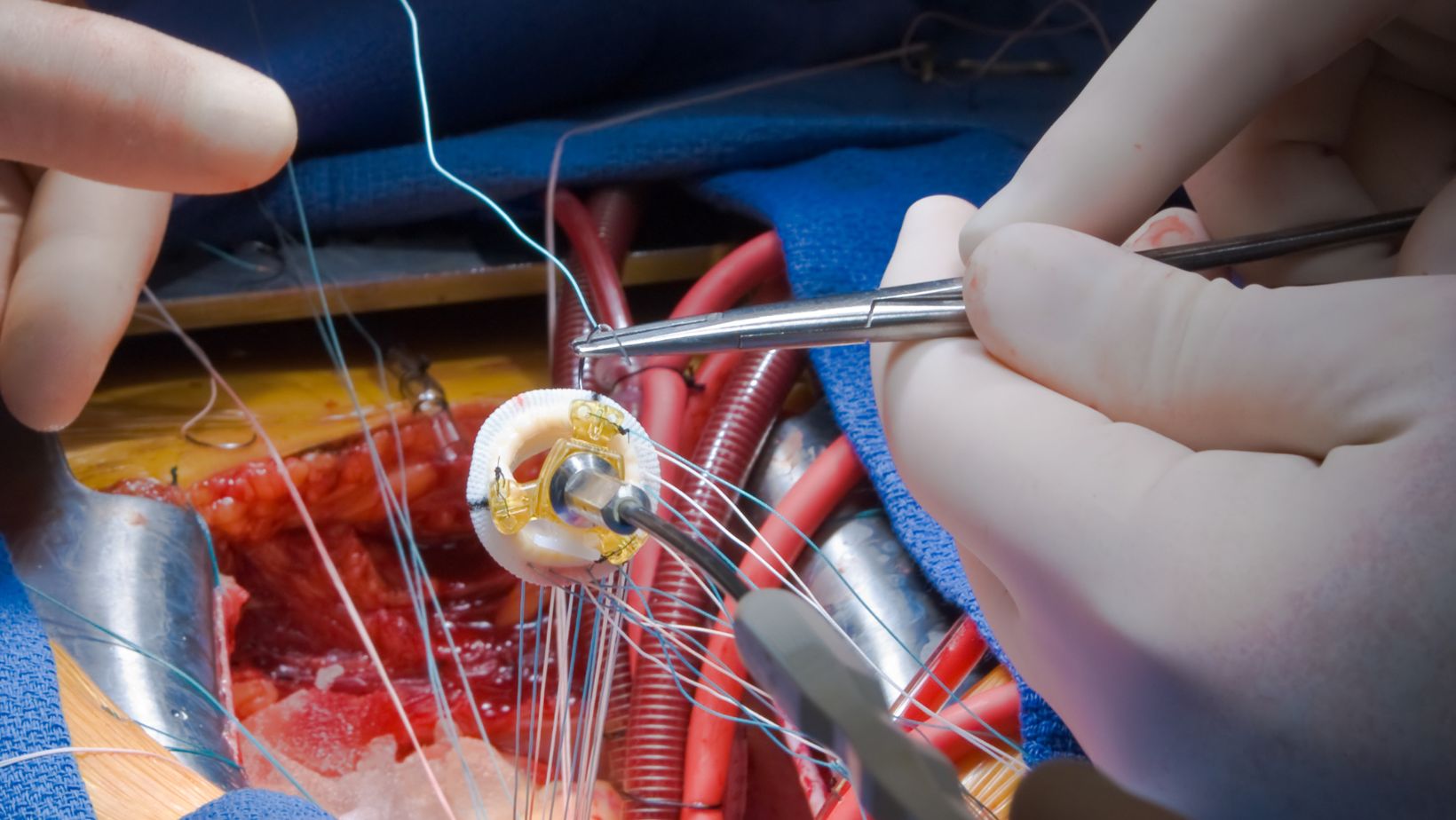
Closed heart surgery with a small incision in the chest is a method that has entered everyday surgical life through an incision of 3-4 cm in length. Patients prefer it due to the small size of the incision, but, as always, the final decision depends on the doctor’s testimony. Here, the heart valve is completely removed and replaced with a mechanical or biological valve. The operation time is quite short. This takes approximately 1.5-2 hours. During this process, the old calcined coating is removed and all lime is cleaned and removed. The valve is stitched and installed in place. Therefore, a heart-lung machine is needed. With the closed method, only 3-4 cm of bone is cut, depending on where it is performed.
Method 3 – TAVI
The TAVI method is a method developed in recent years. With this method, the chest does not open, and the aortic valve is replaced with a biological valve inserted through the groin. Additionally, in the TAVI method, a biological aortic valve placed in a stent is attached to patients without the need for a heart and lung machine. The patient’s own defective original aortic valve is not removed, as in surgical methods, but is left inside by compressing it with the stented valve. The biggest advantage of this method is that it is a completely closed method and there is no need for a heart-lung machine. However, this method also has serious disadvantages. With the TAVI method, only a biological aortic valve can be installed. The biological valve may become unusable over a long period (denaturation) and may have to be replaced again. Some complications may occur with the TAVI method. For example, in cases where the valve has slipped or does not fit properly, emergency or elective open-heart surgery may be required. Even if the valve fits perfectly, blood may leak from the edge of the valve, which we call a “paravalvular leak.” In this case, patients may undergo open heart surgery in the future. With TAVI, the original and calcified valve is usually ruptured and a new valve is placed on top of the old one. This rupture is more likely to cause blood clots (emboli) in the brain vessels than with other surgical techniques.
With TAVI, a new valve is inserted through the groin using a catheter to the heart. For this reason, the inguinal and abdominal veins must be of appropriate diameter and not calcified. Otherwise, these vessels may rupture, and in emergencies, it is necessary to open the abdominal cavity and restore this vessel. During the TAVI procedure, cardiac angiographic monitoring is performed. Even in the most elite centers, the mortality rate from the TAVI procedure is approximately 10%.
Patients undergoing aortic valve surgery should carefully evaluate all of these options and make their own decisions accordingly. In all 3 methods, the experience of the person performing the procedure and the team is very important. Hospital infrastructure, the TAVI surgical team’s availability, and the team’s experience play an important role in the procedure’s outcome. Although each method has its advantages and disadvantages, aortic valve stenosis (stenosis) or incompetence is a serious condition and requires treatment. Otherwise, patients may be subject to heart failure, embolism, and death.
When should surgery be performed for aortic valve disease?
In cases where the aortic valve allows blood to flow backward, there is severe stenosis, calcification, impaired valve function, or all of them occur simultaneously, serious heart rhythm disturbances and the risk of blood clots occur, and the patient’s life is at risk. These patients are urgently evaluated and surgery is indicated.

Unlocking Growth: 4 Strategies for Fintech Marketing Agencies

What Does the Future of Climate Mean for Water Damage & Mold Removal

7 Benefits of Having a Dedicated Legal Team for Your Company

Make Your Life Easier as an Indian Expat in the USA

4 Ways to Earn Money With Little to No Upfront Costs

Benefits of Registering a Forex Broker in Saint Lucia

What Are the Benefits of Buying an FSA-Eligible Mattress?

6 Benefits of Regular Check-ups at Franklin Vet Clinic

5 Tips for Overcoming Addiction

Your Guide to QA Courses for Career Advancement
-

 Personal Finance5 months ago
How Do I Find My UCAS ID Number?
-
Success5 years ago
Consistency: The Key Ingredient to Success
-
Uncategorized5 months ago
What Does Conditionally Approved Mean For An Apartment?
-
Motivation2 years ago
How To Become a More Organized Person?
-
Others4 years ago
Work Health and Safety: 8 Reasons to Maintain a Clutter-free Office
-
Entrepreneurs3 years ago
Why Diversity is Key in Business Marketing
-
HK Pools5 months ago
The HK Pools Forum Comunity Jos Markotop 2D Warna Kuning – A Great Way to Stay Connected
-
Sport1 year ago
What Makes Soccer Betting So Great?