
Understanding the Visual Field Test Machine – Why Is It Important
The human eye is a masterpiece of an organ that can do marvelous things like perceive depth, movement, and become adapted to light and shadows. Ophthalmologists and optometrists have to look past the central vision to provide meaningful insight on the health of our eyes. They also need to examine what we perceive at the edges—our peripheral vision. This is where a visual field test machine comes in handy.
These machines play the key roles in the diagnosing, management and monitoring various eye and neurological disorders. As diagnostic technology has improved, the visual field test machine has also become more accurate, quicker, and comfortable to both the practitioner and the patient. So, what is the role of this machine and why is it so significant?
What is Visual Field Test?
A visual field test involves analyzing the whole range of vision of an individual including central as well as peripheral. It uses how far and how clearly one can see all around without moving his or her eyes further to determine the visual field in all directions. This is essential in identification of the blind areas or areas where the visibility can be impaired or lacking.
This test is particularly vital as most eye conditions start attacking the peripheral vision instead of the central one which implicates that patients may not notice that there is something wrong until the condition has advanced to an extreme level.
How the Machine for Visual Field Testing Operates
The current visual field test equipment (https://www.optometrytimes.com/view/visual-field-testing-undergoes-modern-transformation-via-wearable-technology) often known as a perimeter, measures a person’s ability to detect light at different angles and intensities using controlled stimuli.
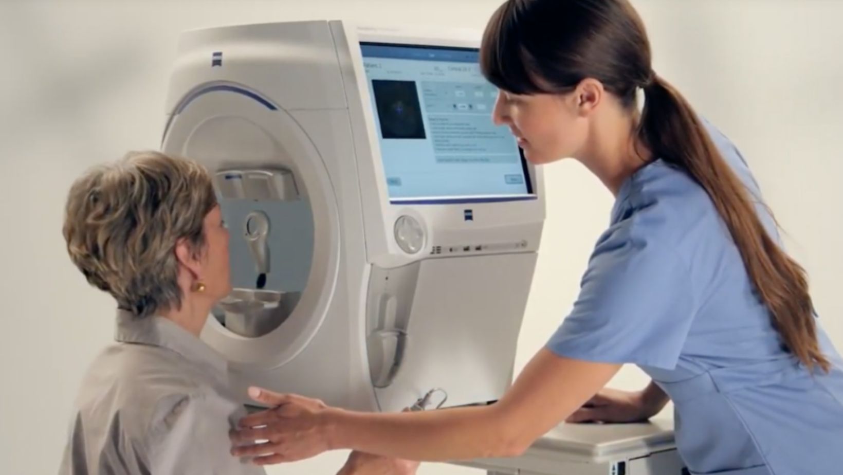
Automated static perimeters, like the Humphrey Field Analyzer (HFA), are the most often used kind. Here’s how the test usually goes:
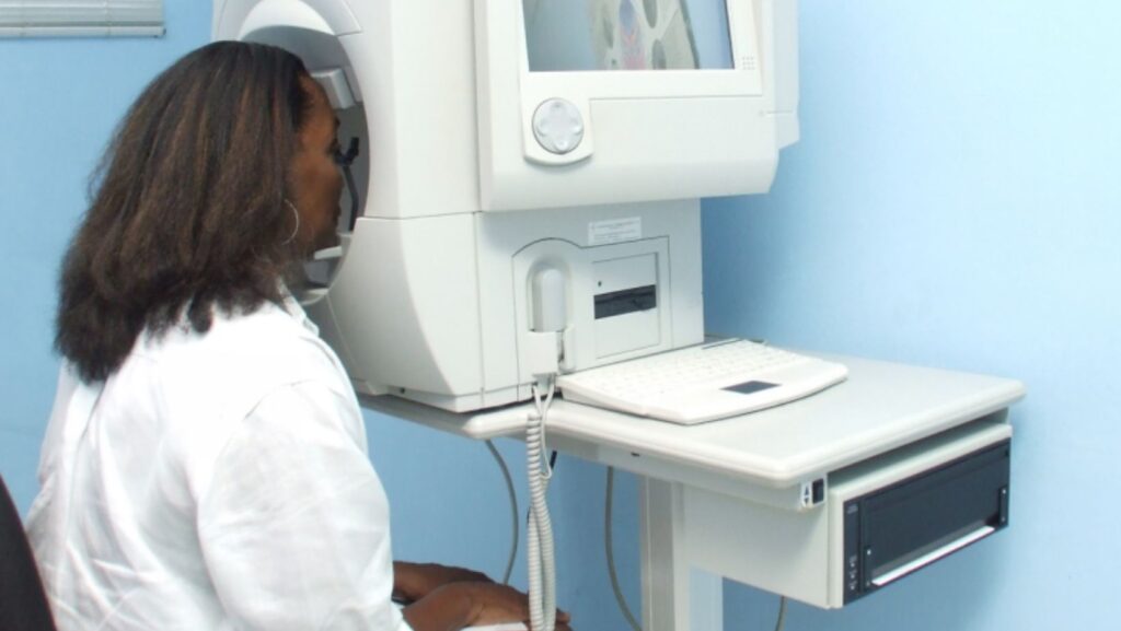
First, the patient sits in front of a concave dome. With one eye shielded, the other concentrates on a stationary focal point. The field of view is filled with sporadic bursts of light. Then, the patient presses a button whenever they see a flash.
The device creates a “visual field map” by charting the regions where the patient reacts and those that do not. Reduced or lost vision is indicated by darker spots on the map.
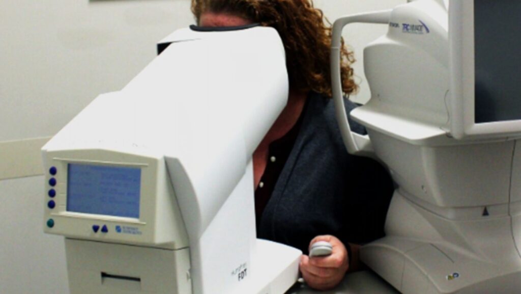
Additionally, some devices provide kinetic perimetry, which measures the outermost range of a person’s field of vision using moving light targets. Others are set up for testing with frequency doubling technology (FDT), which employs low spatial frequency stimuli to identify retinal ganglion cell function loss early on.
The Significance of Visual Field Testing
Visual field testing is essential for both monitoring and diagnosing a wide range of conditions:
Glaucoma
One of the main applications for a visual field test machine is this. Loss of peripheral vision is frequently the first sign of glaucoma, which damages the optic nerve gradually. The device tracks the course of the disease over time and aids in the early detection of damage.
Disorders of the Nervous System
The brain’s visual circuits can be affected by diseases like multiple sclerosis, brain tumors, and strokes. The test assists in detecting patterns of vision loss that point to potential sites of neurological injury.
Retinal Diseases
Both central and peripheral vision can be impacted by conditions like diabetic retinopathy and retinitis pigmentosa. For the purpose of diagnosis and treatment, mapping out these changes is essential.
Medication Monitoring
Retinal toxicity can result from some drugs, such as hydroxychloroquine, which is used to treat autoimmune diseases. To identify early vision abnormalities before irreversible damage occurs, routine visual field testing is crucial.
Visual Field Test Machine Developments
In this field, technology has advanced quickly. The visual field test instruments of today are made to be more precise, effective, and comfortable.
Algorithms in contemporary machines shorten test times without compromising precision. Test validity has increased since devices can now identify when a patient is not paying attention or is clicking excessively. Testing can be done in non-traditional settings, such as home visits or rural clinics, thanks to portable and small perimeters.
To expedite diagnosis and treatment, test results can be electronically transferred to a patient’s electronic medical record. In order to improve early diagnosis rates and assist physicians in identifying minor changes over time, some more recent models even integrate AI-driven analysis.
The Patient Experience
A visual field exam is usually painless and straightforward for patients, although it does call for cooperation and focus. Results may be impacted by blinking, exhaustion, or a lack of comprehension. For this reason, skilled technicians frequently conduct practice tests and provide a thorough explanation of the procedure before starting.
Design enhancements such as ergonomic sitting, adjustable chin rests, and improved illumination have also lessened testing-related discomfort and tiredness.
Pediatric-friendly equipment and modified methods make the test more accessible for kids and people with impairments.
How Frequently Are Visual Field Tests Needed?
The risk profile of the individual determines how frequently they should be tested. Healthy people might only need to be screened once or twice during regular eye exams. To track the disease’s progression, glaucoma patients may require testing every three to six months. It is frequently advised to conduct routine testing for medication monitoring every six to twelve months.
To customize the testing regimen for each patient, doctors use clinical complaints, intraocular pressure readings, optic nerve imaging, and visual field data.
Interpreting the Results
Several kinds of maps and charts are produced by the visual field machine. The difference between the patient’s reactions and those of a healthy population is displayed on the total deviation map. The pattern deviation map emphasizes localized damage while adjusting for overall visual loss brought on by cataracts or small pupils.
In particular, the Glaucoma Hemifield Test (GHT) examines patterns that are common in glaucomatous damage. Progression analysis shows the evolution of the field over time.
Physicians use these findings to determine if a patient’s health is stable, getting better, or getting worse, as well as whether to begin, intensify, or modify treatment.

Singapore’s Favorite Casino Games: What Makes Them So Popular in 2025

6 Common Lab Tools Used in Single-Cell Research

How Food Trend Predictions Are Shaping a Healthier, More Sustainable Future

How social casinos are redefining gaming in the digital age

Effective Sports Gear for Every Workout Routine

Why Custom Made Mylar Bags Work: 15 Key Stats That Prove Their Power

Empowering Dental Practices Through Purpose-Driven Website Design

Derma Stamp vs Dermaroller: Which Works Better

Where to Purchase Apple Gift Card: A Complete Guide for Gamers and Everyday Users

Minimally Invasive Techniques in Colorectal Surgery: Advancements and Challenges with Insights from Dr. Omar Marar
-

 Success7 years ago
Consistency: The Key Ingredient to Success
-
Personal Finance2 years ago
What Does Conditionally Approved Mean For An Apartment?
-
HK Pools2 years ago
The HK Pools Forum Comunity Jos Markotop 2D Warna Kuning – A Great Way to Stay Connected
-
Personal Finance2 years ago
What Letter Grade Is 16 Out Of 20? |
-
 Interesting Facts1 month ago
Introduction: Is Flirt.com The Website I Know I Can trust?
-
Gift Card Facts2 years ago
Do Trader Joe’s Gift Cards Expire? Find Out the Truth Here!
-
Latest News2 years ago
Isekai Kita no De Special Skill – An In-Depth Analysis and Insights
-
Gift Card Facts2 years ago
How to Check and Manage Your Crumbl Cookie Gift Card Balance